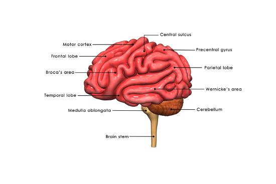
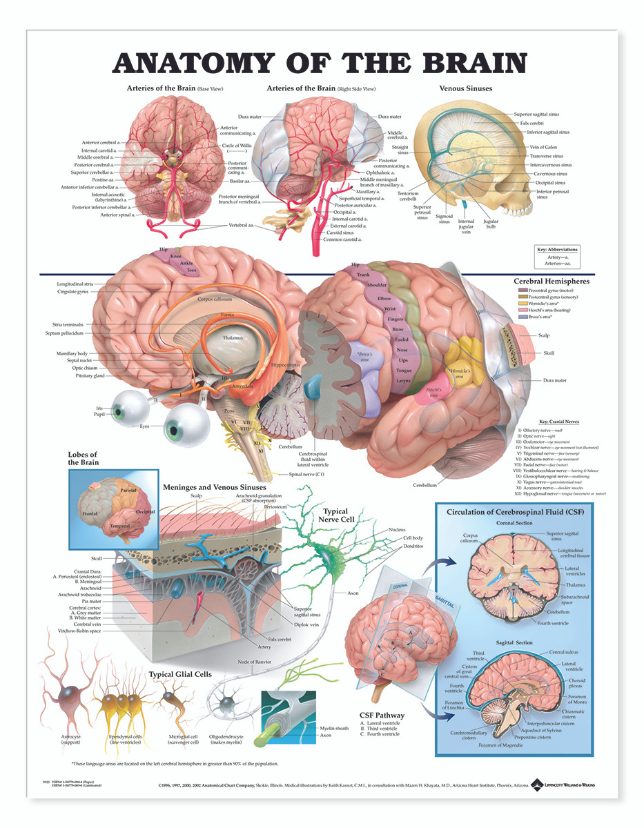
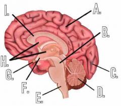
39 labels of the human brain
Human Brain Model Anatomy 4-Part Model of Brain w/Labels & Display Base ... Human Brain Model Anatomy 4-Part Model of Brain w/Labels & Display Base Color-Coded Life Size Human Brain Anatomical Model for Science Classroom Study Display : Amazon.ca: Industrial & Scientific Anatomy of the Brain: Structures and Their Function - ThoughtCo Brain Divisions . The forebrain is the division of the brain that is responsible for a variety of functions including receiving and processing sensory information, thinking, perceiving, producing and understanding language, and controlling motor function. There are two major divisions of forebrain: the diencephalon and the telencephalon. The diencephalon contains structures such as the ...
Diagram Of Brain with their Labelings and Detailed Explanation A well-labelled diagram of a human brain is given below for further reference. Structure And Function Of The Human Brain Parts Of The Human Brain The human brain is divided into three main parts: Forebrain. Midbrain. Hindbrain. These three main parts comprises many small parts. Forebrain The forebrain is also called as Prosencephalon.

Labels of the human brain
2,782 Labeled brain anatomy Images, Stock Photos & Vectors - Shutterstock 2,783 labeled brain anatomy stock photos, vectors, and illustrations are available royalty-free. See labeled brain anatomy stock video clips Image type Orientation People Artists Sort by Popular Healthcare and Medical Anatomy Icons and Graphics human brain brain organ medicine cerebral cortex cerebellum human body Next of 28 Amazon.com: brain model labeled 1-16 of 503 results for "brain model labeled" RESULTS Amazon's Choice Learning Resources Cross-section Brain Model - 2 Pieces, Ages 7+ Brain Anatomy Model, Brain Functions Model, Human Anatomy for Kids, Foam Brain Model 1,393 $18 88 $19.99 Get it as soon as Wed, Mar 30 FREE Shipping on orders over $25 shipped by Amazon More Buying Choices Human Brain - Structure, Diagram, Parts Of Human Brain The thalamus is a small structure, located right above the brain stem responsible for relaying sensory information from the sense organs. It is also responsible for transmitting motor information for movement and coordination. Thalamus is found in the limbic system within the cerebrum.
Labels of the human brain. Nervous System - Label the Brain - TheInspiredInstructor.com This brain part controls balance, movement, and coordination. (11) This brain part controls involuntary actions such as breathing, heartbeats, and digestion. (12) This part of the nervous system moves messages between the brain and the body. (13) This part of the cerebrum interprets and sorts information from the senses. (14) Labeled Diagrams of the Human Brain You'll Want to Copy Now The central core consists of the thalamus, pons, cerebellum, reticular formation and medulla. These five regions are the central areas that regulate breathing, pulse, arousal, balance, sleep and early stages of processing sensory information. The thalamus interprets the sensory information and helps determine what is good and bad. The Human Brain Atlas at Michigan State University The Human Brain Atlas Keith D. Sudheimer, Brian M. Winn, Garrett M. Kerndt, Jay M. Shoaps, Kristina K. Davis, Archibald J. Fobbs Jr., and John I. Johnson Radiology Department, Communications Technology Laboratory, and College of Human Medicine, Michigan State University; National Museum of Health and Medicine, Armed Forces Institute of Pathology Frontiers | 101 Labeled Brain Images and a Consistent Human Cortical ... Labeling the macroscopic anatomy of the human brain is instrumental in educating biologists and clinicians, visualizing biomedical data, localizing brain data for identification and comparison, and perhaps most importantly, subdividing brain data for analysis.
Labeled Parts Of The Brain Illustrations, Royalty-Free Vector ... - iStock Labeled educational bloodstream example Anatomy of Nerves of Body and Head Big collection business, education, online training, marketing... Silhouette brain isolated Silhouette of a brain isolated on a white background consisting of round particles Big collection business, education, online training, marketing... Anatomy of Veins and Arteries Automated Talairach Atlas labels for functional brain mapping An automated coordinate‐based system to retrieve brain labels from the 1988 Talairach Atlas, called the Talairach Daemon (TD), was previously introduced ... This is exemplified by the broad use of the 1988 Talairach atlas by the human brain mapping community [Steinmetz et al., 1989; Fox 1995]. Subcortical structures such as thalamus, caudate ... Brain Anatomy and How the Brain Works - Hopkins Medicine Each brain hemisphere (parts of the cerebrum) has four sections, called lobes: frontal, parietal, temporal and occipital. Each lobe controls specific functions. Frontal lobe. The largest lobe of the brain, located in the front of the head, the frontal lobe is involved in personality characteristics, decision-making and movement. Amazon.com: XINDAM 3D Human Brain with Labels Anatomical Model ... This item: XINDAM 3D Human Brain with Labels Anatomical Model Paperweight (Laser Etched) in Crystal Glass Ball Science Gift (Included LED Base) $66.99 Brain 11 Ounce Ceramic Coffee Mug (WC462M) $16.98 Anatomic Brain Specimen Coasters (Set of 10) - Neuroscience Gifts, Gifts for Medical Student Gifts Brain Decor Human Anatomy Gifts
Main Parts of the Human Brain and Subdivisions of Human Brain Parts Human encephalon resembles, in structure and function, with that of the other vertebrates. The scientists reveal that parts of the human brain are Forebrain, Midbrain & Hindbrain and the related structures that collectively act as a single highly specialized unit. These parts work in coordination and perform different functions of brain. More ... the brain with labels brain labels human inside labeled Lateral View Of The Brain Centered At The Level Of The Intraparietal brain sulcus lateral intraparietal neuroanatomy centered level What Are The 4 Main Types Of Electrical Injury? - Pat Labels electrical types labeled human brain mri brain anatomy normal radiology imaging google human callosum corpus atlas system knee. 33 Human Brain With Label - Labels Database 2020 otrasteel.blogspot.com. brain label human diagram labels drawing labeled draw parts medulla neat ii anatomy drawings psychology cerebellum paintingvalley Brain Basics: Know Your Brain | National Institute of Neurological ... The brain can be divided into three basic units: the forebrain, the midbrain, and the hindbrain. The hindbrain includes the upper part of the spinal cord, the brain stem, and a wrinkled ball of tissue called the cerebellum ( 1 ). The hindbrain controls the body's vital functions such as respiration and heart rate.
Labeled Brain Model Diagram | Science Trends The cerebrum is the largest and most complex portion of the human brain. The cerebrum's function is to control our actions and thoughts, either conscious or unconscious, and responses to stimuli. The cerebrum itself is typically divided into four different lobes: the temporal lobe, the parietal lobe, the occipital lobe, and the frontal lobe.
Solved Label the structures and lobes of the human brain by - Chegg Label the structures and lobes of the human brain by clicking and dragging the labels to the correct location. <--Anterior Posterior --> Precentral gyrus Temporal lobe Parieto-occipital sulcus Parietal lobe Lateral sulcus Insula Postcentral gyrus Central sulcus Occipital lobe Frontal lobe Reset Zoom
File:Brain human normal inferior view with labels en.svg Original upload log []. File:Brain_human_normal_inferior_view.svg licensed with Cc-by-2.5 . 2009-10-13T16:18:05Z Beao 424x505 (209117 Bytes) Replaced right brain half with a clone of left brain half because they look excly the same in the picture.; 2007-09-23T15:14:17Z Ysangkok 424x505 (417241 Bytes) removing credits; 2007-03-03T17:30:01Z Ysangkok 424x505 (417718 Bytes) trying to make it work ...
brain labeled diagram The Human Brain Anatomy - YouTube we have 9 Images about The Human Brain Anatomy - YouTube like Brain And Spinal Cord Diagram Anatomy Chart Of Spinal Cord Labeled, Functions of The Hippocampus Unveiled and also Brain And Spinal Cord Diagram Anatomy Chart Of Spinal Cord Labeled. Read more: The Human Brain Anatomy - YouTube
Human Brain Anatomy - Components of Human Brain with Images Gray & White Matter of Brain: It is one of the amazing brain facts that there are around 100 billion neurons or nerve cells in the brain, the majority of which (about 70%) are found in the cerebral cortex. The nerve cell bodies and axons have graying and whitish appearances, respectively.
The Human Brain | Brain and Cognitive Sciences | MIT OpenCourseWare Image of a cortex with colored labels of the regions resposible for various perceptual and cognitive functions. (Courtesy of the instructor.) Course Description This course surveys the core perceptual and cognitive abilities of the human mind and asks how they are implemented in the brain.
3D Brain This interactive brain model is powered by the Wellcome Trust and developed by Matt Wimsatt and Jack Simpson; reviewed by John Morrison, Patrick Hof, and Edward Lein. Structure descriptions were written by Levi Gadye and Alexis Wnuk and Jane Roskams .
The Human Brain - Visible Body It consists of three structures: the medulla oblongata, the pons, and the midbrain. The medulla oblongata is continuous with the spinal cord and connects to the pons above. Both the medulla and the pons are considered part of the hindbrain. The midbrain, or mesencephalon, connects the pons to the diencephalon and forebrain.
Label the Brain Anatomy Diagram Flashcards | Quizlet Label the Brain Anatomy Diagram. STUDY. Flashcards. Learn. Write. Spell. Test. PLAY. Match. Gravity. Created by. eileenvalverde97. Terms in this set (18) ... Human Anatomy & Physiology 8th Edition Elaine N. Marieb, Katja Hoehn. 873 explanations. Essentials of Human Anatomy & Physiology 10th Edition Elaine N. Marieb.
7 Best Images of Functions Of The Brain Worksheet - Brain Anatomy Diagram Unlabeled, Blank Brain ...
Brain (Human Anatomy): Picture, Function, Parts, Conditions, and More Human Anatomy. The brain is one of the largest and most complex organs in the human body. It is made up of more than 100 billion nerves that communicate in trillions of connections called synapses ...
Human brain - Wikipedia The surface of the brain is folded into ridges ( gyri) and grooves ( sulci ), many of which are named, usually according to their position, such as the frontal gyrus of the frontal lobe or the central sulcus separating the central regions of the hemispheres. There are many small variations in the secondary and tertiary folds. [19]
Human Brain - Structure, Diagram, Parts Of Human Brain The thalamus is a small structure, located right above the brain stem responsible for relaying sensory information from the sense organs. It is also responsible for transmitting motor information for movement and coordination. Thalamus is found in the limbic system within the cerebrum.
Amazon.com: brain model labeled 1-16 of 503 results for "brain model labeled" RESULTS Amazon's Choice Learning Resources Cross-section Brain Model - 2 Pieces, Ages 7+ Brain Anatomy Model, Brain Functions Model, Human Anatomy for Kids, Foam Brain Model 1,393 $18 88 $19.99 Get it as soon as Wed, Mar 30 FREE Shipping on orders over $25 shipped by Amazon More Buying Choices
2,782 Labeled brain anatomy Images, Stock Photos & Vectors - Shutterstock 2,783 labeled brain anatomy stock photos, vectors, and illustrations are available royalty-free. See labeled brain anatomy stock video clips Image type Orientation People Artists Sort by Popular Healthcare and Medical Anatomy Icons and Graphics human brain brain organ medicine cerebral cortex cerebellum human body Next of 28


Post a Comment for "39 labels of the human brain"